Imaging flow of the standard B-mode ultrasound imaging. PRF
5 (655) In stock

.jpg)
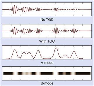
The A, B, M's – Ultrasound Modes Explained

Jong Chul YE, Ph. D., Korea Advanced Institute of Science and Technology, Daejeon, KAIST, Department of Biological Sciences

Physics and Instrumentation in Doppler and B-mode Ultrasonography

Doppler Ultrasound: Principles - ppt video online download

Our network architecture for RF-interpolation.
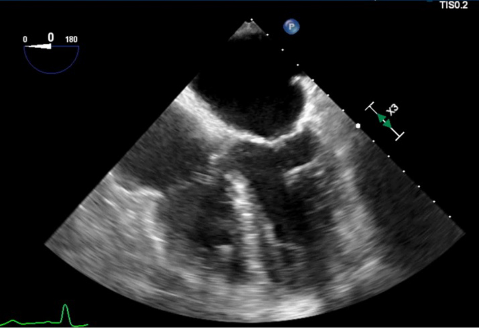
Echocardiography Tutorial - Echocardiographic Modes
Physics and Instrumentation in Doppler and B-mode Ultrasonography

Jaeyoung HUH, Student, Korea Advanced Institute of Science and Technology, Daejeon, KAIST, Department of Bio and Brain Engineering
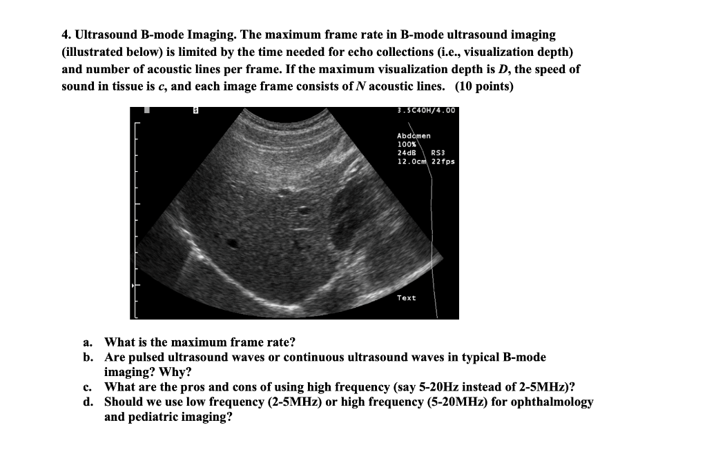
Solved 4. Ultrasound B-mode Imaging. The maximum frame rate

Figure 1 from Deep Learning in RF Sub-sampled B-mode Ultrasound Imaging

Imaging flow of the standard B-mode ultrasound imaging. PRF : pulse

PDF) Deep Learning in RF Sub-sampled B-mode Ultrasound Imaging

New microvascular ultrasound techniques: abdominal applications

Optimizing Image Quality When Evaluating Blood Flow at Doppler US
A) A brightness mode (b-mode) image of the lateral abdominal wall.
Lensing B-modes in the Cosmic Microwave Background polarization
Imaging flow of the standard B-mode ultrasound imaging. PRF : pulse
Versatile Single-Element Ultrasound Imaging Platform using a Water
 Pantalón de vestir en sarga - Beige - MUJER
 calvin klein Classics Women’s Wool 80s Blazer Brown Paisley EUC Size 6 Seinfeld
 SWIM TANK - 120 Madison St, Mamaroneck, New York - Swimming Lessons/Schools - Phone Number - Yelp
 WIZARD OF OZ DOROTHY AND HER SHOES CAPRI LEGGINGS
 How To Clean A Diamond Engagement Ring – Alicia J Diamonds
 Retainer Case, Slim Aligner Case With Vent Holes And Mirror Compatible With Invisalign Night Guard And Mouth Guard